Soft Tissue Anatomy of the Hand Biology Diagrams
Soft Tissue Anatomy of the Hand Biology Diagrams File:DIagram of the different types of soft tissue in the body CRUK 037.svg

Rectus Capitis Muscle Sternocleidomastoid Muscle Palantine Tonsil Soft Palate Retromandibular Vein Maxillary Tuberosity Vertebral Artery in Transverse Foramin Download scientific diagram | Soft tissue anatomy from publication: Soft Tissue Modelling from 3D Scanned Data | Human body 3D scanners are becoming a mature technology that generates accurate Start studying Soft Tissue Anatomy. Learn vocabulary, terms, and more with flashcards, games, and other study tools.

Soft Tissue Anatomy Diagram Biology Diagrams
Supportive connective tissue—bone and cartilage—provide structure and strength to the body and protect soft tissues. A few distinct cell types and densely packed fibers in a matrix characterize these tissues. Download scientific diagram | Anatomy of skin and soft tissue structures and layers commonly involved with various infectious processes. from publication: Treatment of Complicated Skin and Soft Traditionally, the word fascia was used primarily by surgeons to describe the dissectible tissue seen in the body encasing other organs, muscles, and bones. Recently, the definition has been broadened to include all collagenous based soft tissues in the body, including cells that create and maintain the extracellular matrix.
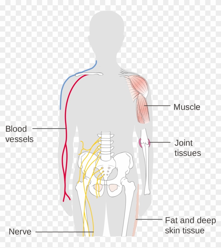
Figure 4.1.2 shows the types of tissues and organs associated with each of the three germ layers. Note that epithelial tissue originates in all three layers, whereas nervous tissue derives primarily from the ectoderm and muscle tissue derives from the mesoderm.
